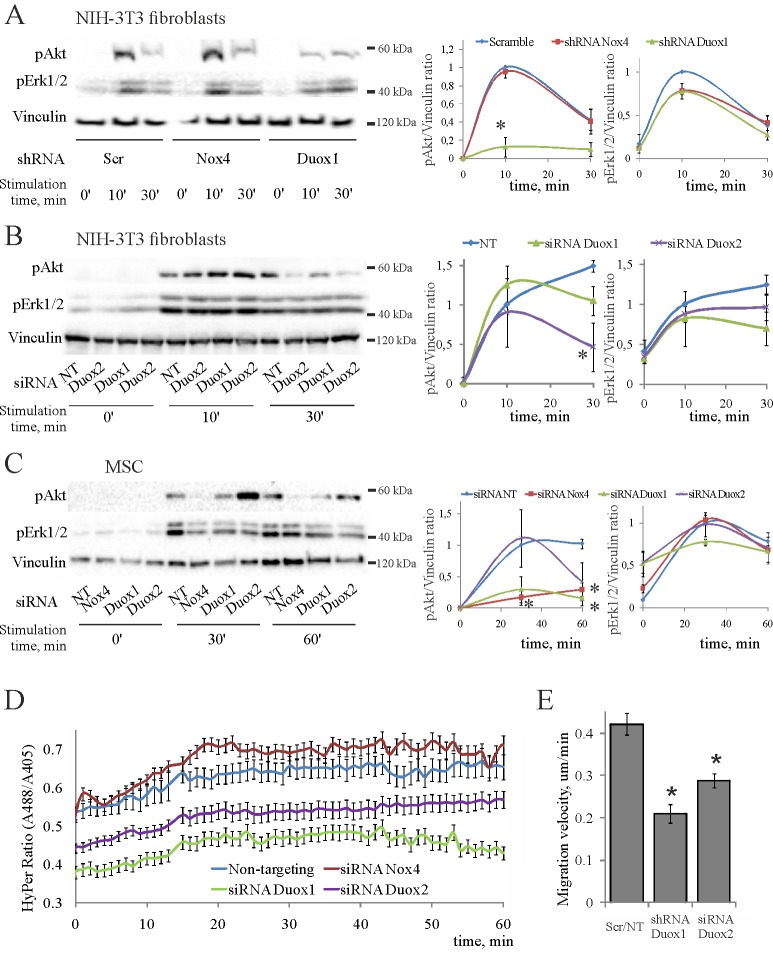
Fig 6. Duox1/2 and Nox4 mediate the PDGF-induced responses in mesenchymal cells.
Representative blots are shown on the left, statistics are shown on the right. The data are normalized to 10 min of stimulation in scrambled/NT controls for fibroblasts and 30 min of stimulation for MSC, which have therefore no error bars; (*) p < 0.05 as compared to scrambe (scr) or non-targeting (NT) controls from 2–4 independent experiments. (A) PDGF-induced phosphorylation of PKB/Akt and Erk1/2 in 3T3 fibroblasts stably expressing the indicated shRNAs. (B) PDGF-induced phosphorylation of PKB/Akt and Erk1/2 in 3T3 fibroblasts transiently transfected by indicated siRNAs. (C) PDGF-induced phosphorylation of PKB/Akt and Erk1/2 in MSC transiently transfected by indicated siRNAs. (D) Kinetics of cytoplasmic H2O2 accumulation in 3T3 fibroblasts pre-treated by indicated siRNAs; PDGF added at 0 min. (E) Effects of Duox1/2 silencing on speed of 3T3 fibroblast migration. Shown are the results of shRNA- and siRNA-mediated silencing of Duox1/2 compared, respectively, to scramble and NT controls, which moved with identical speeds.

