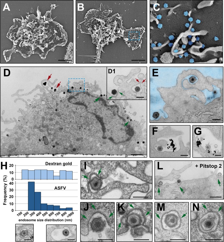
Fig 2. ASFV endocytosis at the ultrastructural level.
Mock (A) and ASFV-infected (B and inset C) (MOI 100) swine macrophages were fixed at 20 mpi and analyzed by field emission scanning EM. The characteristic cell membrane protrusions of monocyte/macrophage lineage can be visualized in both conditions. ASFV particles (depicted in blue in inset C) are detected on flat membrane areas and also in the interstices of the membrane evaginations. D-E, I-J, L-M) ASFV-infected macrophages (MOI 100) were examined at 10 mpi by transmission EM after fixation. Panel D shows a side view of an infected macrophage. Note that ASFV particles seem to be endocytosed by macropinocytic-like membrane protrusions (red arrows) as well as by membrane invaginations characteristic of clathrin-dependent endocytosis (green arrows). Inset D1 shows a detail of panel D at higher magnification. E shows a virion apparently engulfed by macropinocytosis. F-G) Macrophages were incubated with both ASFV and dextran-coated 30-nm gold particles for 7 min at 37°C. F shows macropinocytic-like uptake of both ASFV and dextran-gold particles and G shows colocalization at a putative macropinosome. H) Size distribution histogram of endosomes containing either dextran-coated 30-nm gold particles (upper) or ASFV particles (lower) after 7 min of incubation. I-J, L-M) ASFV uptake by clathrin-coated pits (I, J and K) and coated vesicles (M and N). The clathrin coats are indicated by arrows. L) ASFV-infected macrophages were incubated for 15 min with 15 μM Pitstop 2. Arrows indicate virions at coated pits. Bars, 200 nm except for panels A and B (5 μm), D (1 μm) and L (500 nm).

