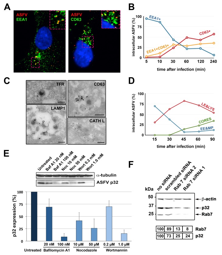
Fig 4. Transport of incoming ASFV along the endocytic pathway.
A-B) ASFV-infected Vero cells (MOI 10) were analyzed at the indicated times by immunofluorescence detection of viral protein p17, early endosome/macropinosome marker EEA1 and late endosome/lysosome marker CD63. A) Colocalization of incoming viruses (red) with EEA1+ vesicles (green) at 10 mpi (left) or with CD63+ vesicles (green) at 30 mpi (right). B) Quantification of ASFV particles colocalizing with either EEA1, CD63 or both markers at the indicated times. C) Immunoelectron microscopy of incoming ASFV particles within endocytic vesicles labeled for early endosomal marker TFR, late endosomal/lysosomal markers CD63 and LAMP1, and lysosomal cathepsin L (CATH L). Immunogold labeling was performed on thawed cryosections of ASFV-infected cells using the indicated antibodies and protein A-gold (15 nm) conjugates. Bars, 200 nm. D) Transport of incoming ASFV in swine macrophages. Infected macrophages (MOI 100) were processed by EM at the indicated times. For each time, intracellular particles were classified and quantified according to their presence within early endosomes and macropinosomes (EE&MP), late endosomes and lysosomes (LE&LYS) or at the cytosol (CORES). E) ASFV-infected macrophages (E70, MOI 5) were treated for 2.5 h with inhibitors of endosome maturation (Baf A1, nocodazole and wortmannin) and analyzed for expression of viral protein p32 by immunoblotting (upper panel). Graphic shows percentage of p32 expression (mean ± SD from triplicate samples) referred to control, non-treated cells. F) COS-1 cells were transfected with two different siRNAs (1 and 2) targeted to Rab7, a scrambled siRNA or not treated (no siRNA). Then, cells were infected with ASFV (MOI 5) for 5 h. Expression of proteins p32, Rab7 and ß-actin was analyzed by immunoblotting. Numbers under the blot image indicated normalized expression levels for Rab7 and p32.

