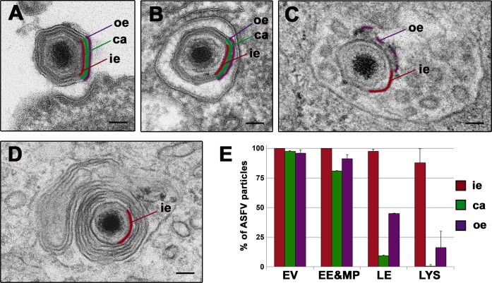
Fig 5. ASFV disassembly in swine macrophages.
Macrophages were infected with ASFV (MOI 100) for 10, 15, 30, 45, 60 and 90 min and processed by EM. A-D) Selected EM images of the endosomal transport and the disassembly process undergone by ASFV. After endocytosis, incoming extracellular particles (A) are first detected (10–15 mpi) inside early endosomes (B) or macropinosomes keeping their structure nearly intact. Later (30–45 mpi), particles are predominantly found at multivesicular late endosomes (C), where a significant proportion loses their protein capsid (depicted in green) and the outer envelope (purple). Finally (45 mpi onwards), a fraction of particles reach lysosome-like structures (D) where most of them appear as a dense cores enwrapped by the inner envelope (red). Bars, 50 nm. E) Quantification of the ASFV disassembly. Incoming virus particles were classified according to their endocytic compartment (EE&MP: early endosomes and macropinosomes; LE: late multivesicular endosomes, LYS: lysosomes) and then, according to their layer content (ie: inner envelope, ca: capsid, oe: outer envelope). Data are expressed as percentages (mean ± deviation of duplicate experiments) of particles with a given domain inside each compartment. As a reference, extracellular virions (EV) attached to the cell surface were also quantified. More than 100 particles per compartment and experiment were analyzed. Note that the loss of the capsid and the outer envelope occurs essentially at multivesicular late endosomes and lysosome-like vesicles.

