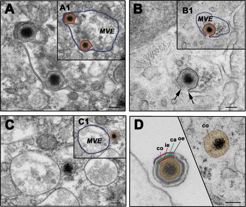
Fig 6. ASFV fuses through the inner envelope at late multivesicular endosomes.
ASFV-infected swine macrophages (MOI 100) were analyzed by EM at 90 mpi. A-D) Representative EM images of the fusion process. After the loss of protein capsid and outer viral membrane, the inner viral envelope becomes exposed, interacting with the luminal face of the limiting membrane of multivesicular late endosomes (MVE) (A). Then, the inner envelope fuses with the endosomal membrane (B) and delivers naked cores into the cytosol (C). As a result of the disassembly and fusion events, extracellular particles lose the three domains surrounding the virus core before reaching the cytosol (D). Insets A1, B1 and C1 show lower magnification images of panel A, B and C, respectively. To facilitate the interpretation, the inner viral envelope is depicted in red, the virus core in brown and the endosomal membrane in purple. Bars, 100 nm.

