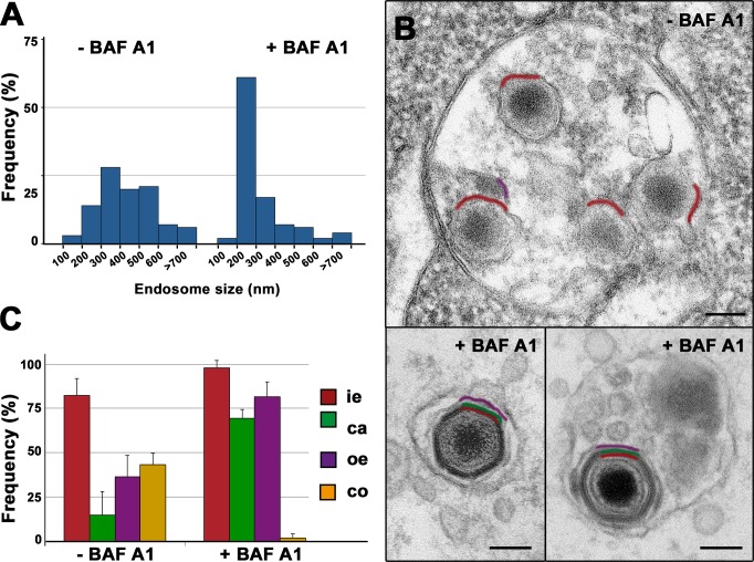
Fig 7. ASFV disassembly and uncoating depends on acidic pH.
Swine macrophages were infected with ASFV for 90 min in the presence or absence of 100 nM Baf A1, an inhibitor of endosomal acidification, and then processed by EM. A) Size distribution of virus-containing endosomes (n>100) in the presence or absence of Baf A1. Note that after drug treatment, the virus-containing endosomes are significantly smaller as a consequence of the impairment of endosome maturation. B) Under these conditions, most ASFV particles appear essentially intact (lower panels). By contrast, most ASFV particles in non-treated cells undergo significant disruption at late endosomes (upper panel). Outer envelope (purple), capsid (green) and inner envelope (red) are indicated. Bars, 100 nm. C) Quantification of Baf A1 effect on virus disruption. Internalized particles from treated (+BAF A1) and nontreated (-BAF A1) cells were quantified according to their layer content (i.e: inner envelope, ca: capsid, oe: outer envelope, co: cytosolic core). Data are expressed as percentages of particles (mean ± SD of triplicate samples) with a given domain for each condition. More than 100 particles per experiment were analyzed. Note that Baf A1 significantly prevents ASFV disassembly and core release. Bars, 100 nm.

