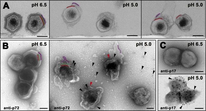
Fig 8. Low pH induces ASFV disruption.
(A) In vitro effect of pH acidification on ASFV structure. Purified virions were exposed to pH 6.5 or 5.0 for 60 min at 37°C and then ultracentrifuged and analyzed by EM after ultrathin sectioning. Note that, at pH 6.5, particles look virtually intact while the particles exposed to acidic pH (5.0) are severely disrupted, displaying evident signals of capsid disassembly as well as disruption and detachment of the outer membrane. Outer envelope (purple), capsid (green) and inner envelope (red) are indicated. (B) Purified virions adsorbed to EM grids were exposed to pH 6.5 or 5.0 for 15 min at 37°C and then immunogold labeled for major capsid protein p72. Note that, at pH 6.5, particles look virtually intact remaining non-accessible for p72 labeling. In contrast, particles exposed to acidic pH (5.0) are severely disrupted. Under these conditions, gold-labeled anti-p72 antibodies (black arrowheads) penetrate the particles through the disrupted outer envelope (red arrowheads), decorating discrete regions where the capsid is still present. Also, p72 labeling is detected on the support film around the particles, as a consequence of capsid disassembly. (C) Purified virions adsorbed to EM grids were exposed to pH 6.5 or 5.0 for 15 min at 37°C and then labeled for major inner envelope protein p17. Note that particles disrupted at pH 5.0 become accessible to anti-p17 labeling. Bars, 100 nm.

