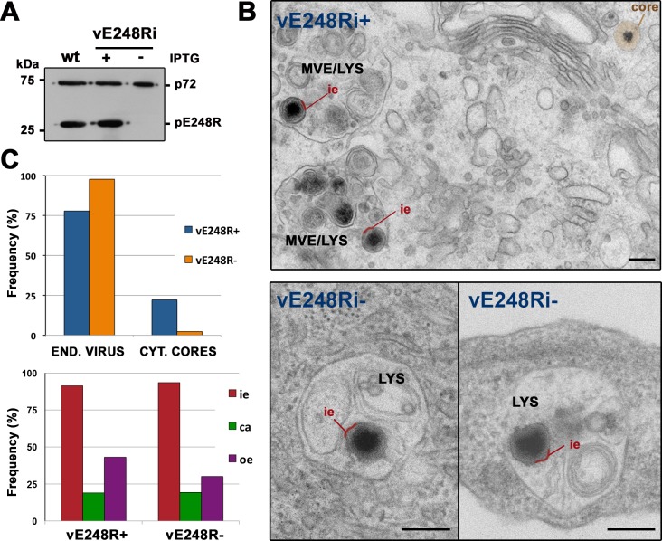
Fig 9. ASFV fusion depends on virus protein pE248R.
A) Percoll-purified ASFV particles were obtained from cells infected with parental (wt) virus or recombinant virus vE248Ri under permissive (+IPTG) and non-permissive (-IPTG) conditions. Virus samples were analyzed by immunoblotting for the presence of capsid protein p72 and pE248R. (B) Vero cells were infected for 2 h with purified recombinant vE248Ri (~ 2 μg for 50,000 cells) grown with (+) or without (-) IPTG and then examined by EM. Disassembly of control vE248R+ (upper panel) and defective vE248R- (lower panels) was similar but core delivery into the cytosol was severely impaired for defective E248R- particles, which accumulated within lysosomes (LYS). To facilitate the interpretation, the inner viral envelope is depicted in red and the cytosolic viral core in brown. MVE (multivesicular endosomes). Bars, 200 nm. C) Quantification of disruption (lower panel) and core delivery (upper panel) of control E248R+ and defective E248R- particles. Endocytosed particles (END. VIRUS) and cytosolic cores (CYT. CORES) of the above experiment were quantified for both conditions and expressed as a percentage of intracellular virus. Also, the endocytosed particles of each condition were classified (lower panel) according to their layer content (i.e: inner envelope, ca: capsid, oe: outer envelope). One representative of two independent experiments is shown.

