
Figure 8.
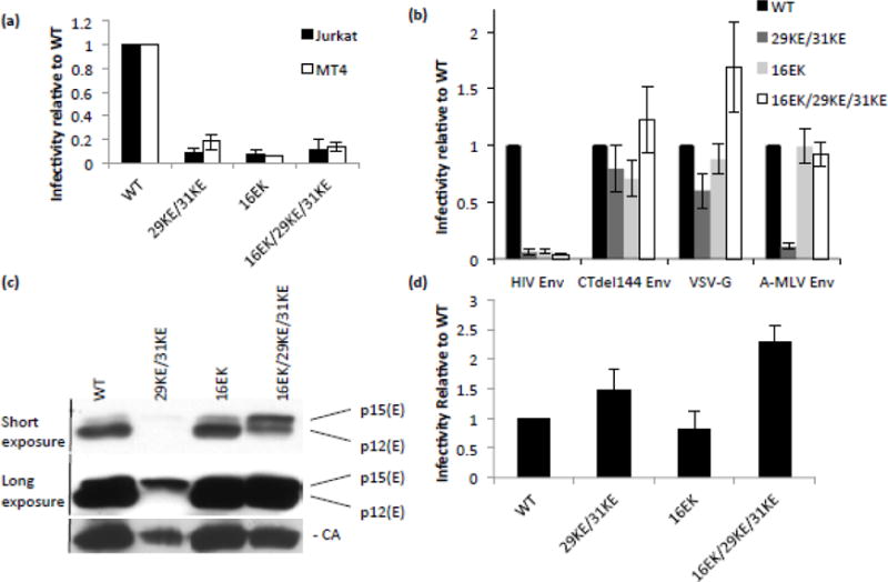
Infectivity and pseudotyping 29/31KE and 16EK. (a) Viruses pseudotyped with VSV-G were generated by transfecting 293T cells with the HIV-1 mutants indicated and a VSV-G expression vector. These viruses were normalized by RT assay and used to infect Jurkat and MT4 T-cell lines. 48 h post-infection virus was collected and RT assays and TZM-bl infections performed. Virus-containing supernatants were used to infect TZM-bl cells; the resulting luciferase signal was normalized to the corresponding RT values to provide a measure of specific infectivity. Means of three independent experiments are plotted, +/− S.E.M. (b) HeLa cells were transfected with Env-deficient clones of the HIV-1 mutants and viral glycoproteins indicated. After 24 h, virus infectivity was determined as in (a). (c) HeLa cells were transfected with Env-deficient clones of the HIV-1 mutants indicated and A-MLV Env. After 48 h, virus was pelleted by ultracentrifugation and analyzed by western blotting. (d) HeLa cells were transfected with Env-deficient clones of the HIV-1 mutants indicated and AMLV p12* Env. After 24 h, virus infectivity was determined as in (a). Means of three independent experiments are plotted +/− S.E.M.