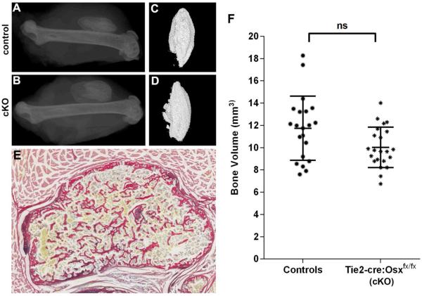
Figure 4.
rhBMP-2 induced ectopic bone formation in Tie2-cre-Osxfx/fx mice and littermate controls. Representative XRs (A, B) and microCT reconstructions (C, D) are shown for the specimens corresponding to the median bone volume for each group. Littermate controls (A, C) were compared to Tie2-cre-Osxfx/fx conditional knockout (cKO) mice (B, D). The bone nodules showed a cortical shell with some trabecular-like elements visualized using Picro Sirius Red/Alcian Blue staining (E). MicroCT quantification revealed no significant difference in bone volume of ectopic rhBMP-2 induced bone with deletion of the Osx gene in Tie2-lineage cells (F).

