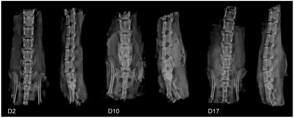
Figure 5.
X-ray images showing the progression of rhBMP-2 induced spine fusion. After implantation of collagen sponges loaded with rhBMP-2, no mineralized bone was seen at D2, but a fusion mass was apparent in all specimens by D10 and D17. Anterior-posterior and lateral views are shown for all time points.

