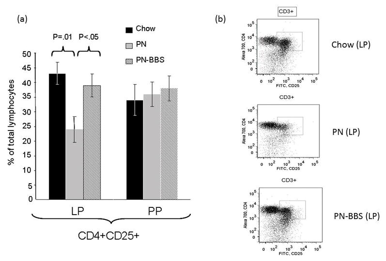
Figure 2. CD4+CD25+ lymphocytes from lamina propria and Peyer’s Patches.

(a)Parenteral nutrition (PN) significantly suppresses the percentage of cells expressing CD4+CD25+ compared to Chow (24% ± 4.4 vs. 43% ± 3.8, p = 0.01). There were no changes in this population in the Peyer’s Patches. Addition of bombesin (BBS) to PN (PN+BBS) significantly increased CD4+CD25+ lymphocytes in the lamina propria compared to PN alone (39% ± 4.4 vs. 24% ± 4.4, p < 0.05) while no changes were seen in the Peyer’s Patches. Values are means ± SE. Flow cytometry gating progressed from the preceding parent population in the order of lymphocytes, CD3+, and then CD4+CD25+ as seen in figure 1. (b)Representative flow cytometry figures from Chow, PN, and PN+BBS demonstrating CD3+ T cells from the parent lymphocyte population. The CD3+ cells were then plotted as CD4+ versus CD25+ and an area (seen in plots as box) of positivity for both markers was determined that was then used for all samples.
