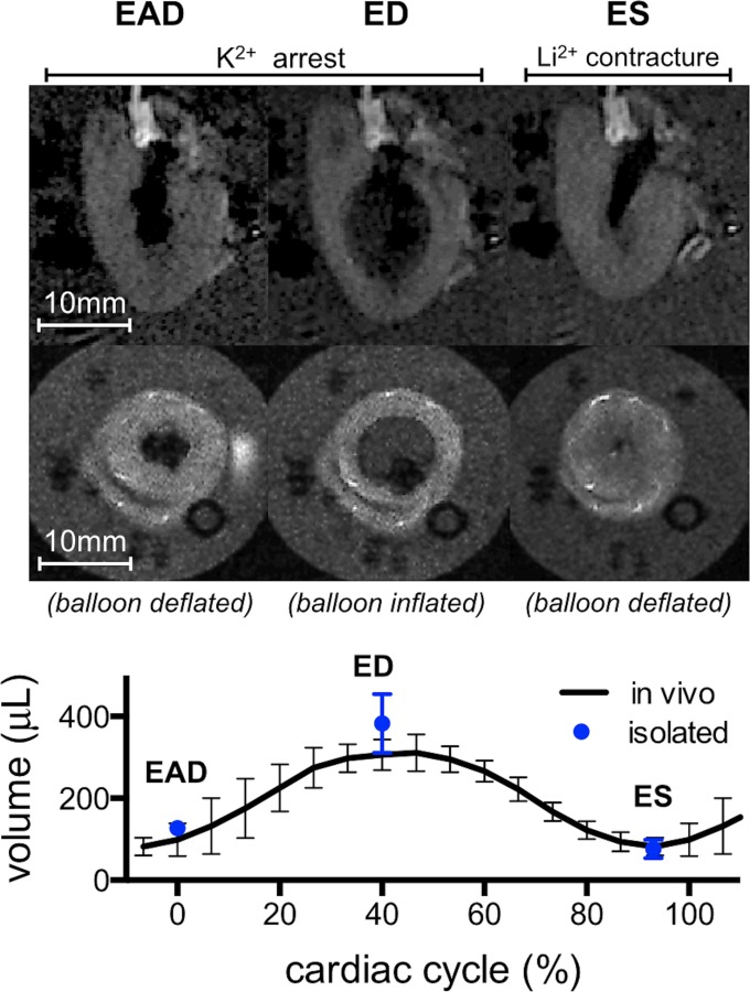
Fig. 4.

Gross morphology of an isolated arrested heart. Long and short axes MRI of the LV (top two rows, see also Fig. 1) revealed morphological alterations consistent with the targeted cardiac states. Compared to EAD, balloon inflation at ED produced volume increase and wall thinning, whereas deflation at ES caused volume decrease and wall thickening. The volumes of the isolated perfused hearts were quantitatively similar to those seen in vivo.
