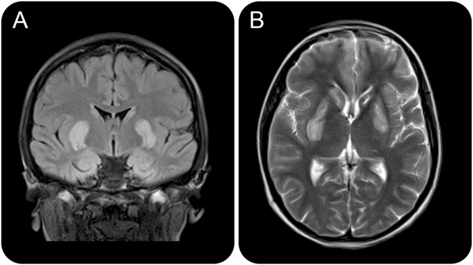
Figure 1. Coronal and axial T2-weighted MRI at presentation.

Coronal fluid-attenuated inversion recovery (A) and axial T2-weighted (B) MRI slices at presentation showing relatively symmetric hyperintensity in the corpora striata, and apparent cystic changes in the left head of caudate.
