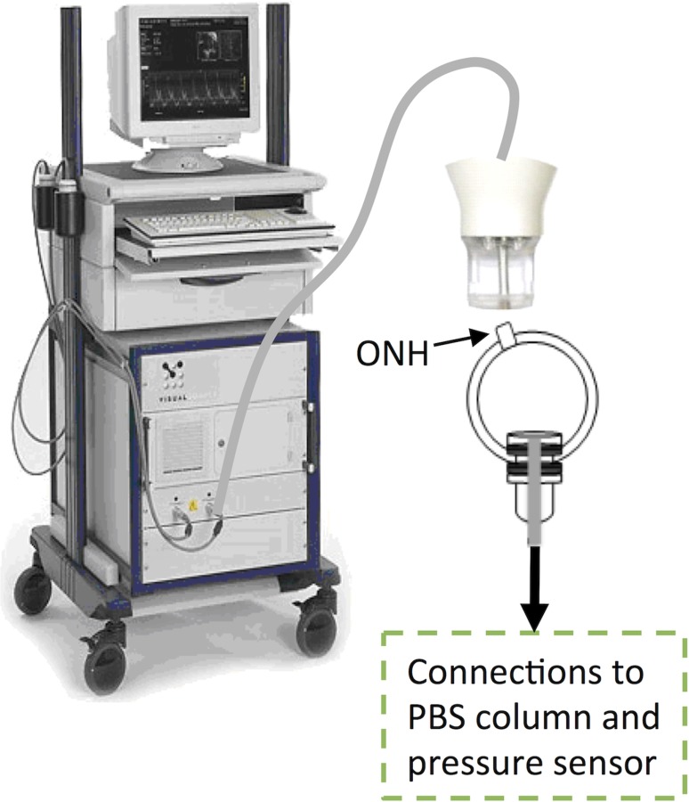
Fig. 1.

Experimental setup of the human donor scleral shell measured with the Vevo 660 high-frequency ultrasound system. ONH: optic nerve head. The shell was clamped near the corneoscleral junction, away from the measured posterior sclera; and immersed in PBS. The ultrasound probe was not in contact with the tissue during scanning.
