Figure 6.
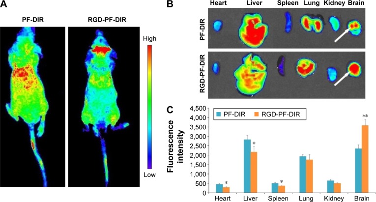
In vivo fluorescence imaging of intracranial U87MG glioma tumor-bearing nude mice 48 hours after intravenous injection of PF-DIR or RGD-PF-DIR (A); representative ex vivo near-infrared fluorescence images of dissected organs of intracranial U87MG glioma tumor-bearing nude mice sacrificed at 48 hours after intravenous injection of PF-DIR or RGD-PF-DIR (B); fluorescence intensity of PF-DIR and RGD-PF-DIR in various organs (C).
Notes: Mean ± standard deviation (n=3). The tumor location is specified with a white arrow. *P<0.05, **P<0.01, compared with the PF-DIR group.
Abbreviations: PF-DIR, dioctadecyl-3,3,3′,3′-tetramethylindotricarbocyanine iodide-loaded Pluronic micelles; RGD-PF-DIR, dioctadecyl-3,3,3′,3′-tetramethylindotricarbocyanine iodide-loaded cyclic arginine-glycine-aspartic acid peptide-decorated Pluronic micelles; U87MG, U87 malignant glioblastoma.

