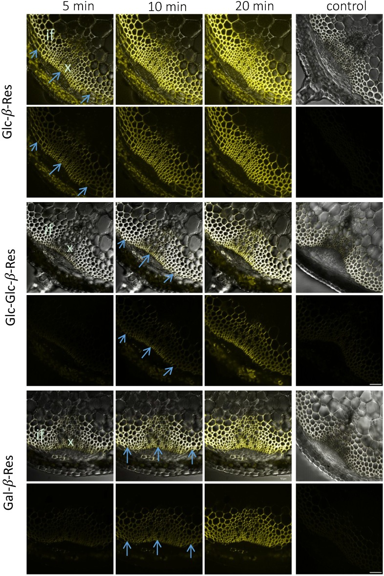
Figure 2.
Distribution of signals in the inflorescence stem sections of Arabidopsis from Glc-β-Res, Glc-Glc-β-Res and Gal-β-Res substrates after incubations for the specified durations. The control sections were boiled for 30 min before incubation with the specified substrate for 20 min. Upper rows represent the transmitted light channel and the superimposed yellow fluorescence channel corresponding to resorufin signals, and the lower rows represent the yellow channel only. Blue arrows show differentiating sclerenchyma cells. if, interfascicular fibers; x, xylem. Bar = 50 μm.

