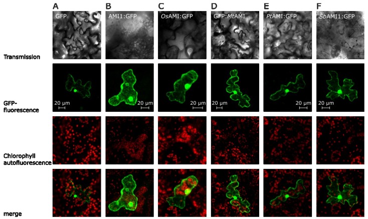
Figure 3.
Localization of amidase GFP fusion proteins in epidermal cells of Arabidopsis thaliana. The images show typical results of confocal laser scanning microscopic studies, using differential emission light filter sets. The upper row provides transmission images, followed by the GFP channel (GFP-fluorescence), showing the fluorescence of the GFP-fluorophore between 500 and 530 nm. Next, the chlorophyll-autofluorescence is shown in a separate channel (650 to 798 nm). In the bottom row, an overlay of the fluorescence channels is depicted. (A) Transformation with an empty GFP-vector (pSP-EGFP), cytoplasmic control; (B) Transformation with an AMI1:GFP construct from A. thaliana [31] (positive control); (C) Transformation of an epidermis cell with the OsAMI1:GFP construct; (D) Transformation with the chimeric GFP:MtAMI construct; (E) Transformation with the PtAMI:GFP vector; (F) Transformation of the SbAMI1:GFP construct. Scale bars for each set of pictures are included in the figure.

