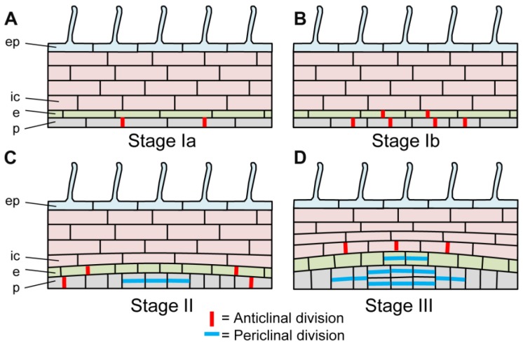
Figure 3.
Schematic representation of the early developmental stages of lateral root formation in Medicago truncatula. Scheme of longitudinal sections in the main root of M. truncatula during LRF, showing the main type of cell divisions but not the precise number of dividing cells. (A). Stage Ia. Anticlinal divisions in the pericycle; (B). Stage Ib. Anticlinal divisions in the endodermis and the pericycle; (C). Stage II. Periclinal divisions in the pericycle and anticlinal divisions in the endodermis and the pericycle; (D). Stage III. Periclinal divisions in the endodermis (two cell layers) and the pericycle (four cell layers), and anticlinal divisions in the inner cortex. p: pericycle; e: endodermis; ic: inner cortex, ep: epidermis. From [61].

