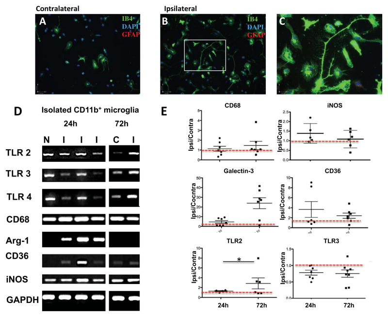
Figure 2. tMCAO changes gene expression of pro- and anti-inflammatory mediators in microglia within injured regions.
A–C. Representative examples of microglial cells isolated 24 hours after tMCAO using CD11b-conjugated magnetic beads and plated on coverslips for 24 hours. Ib4+ cells from contralateral (A) and injured region (B). C. Magnified image in B (white squire). Note the differing morphology of cells from injured and uninjured brain, including the presence of cells with lamellipodia and filopodia. D. Representative RT-PCR examples of pro- and anti-inflammatory genes in isolated microglia. E. Quantification of RT-PCR data in microglia isolated 24 and 72 hours after tMCAO. Data shown as mean±SD for fold change in injured compared to matching contralateral regions. Red dotted line outlines the level in contralateral region. N – naïve, C – contralateral, I – ipsilateral. * - p<0.05.

