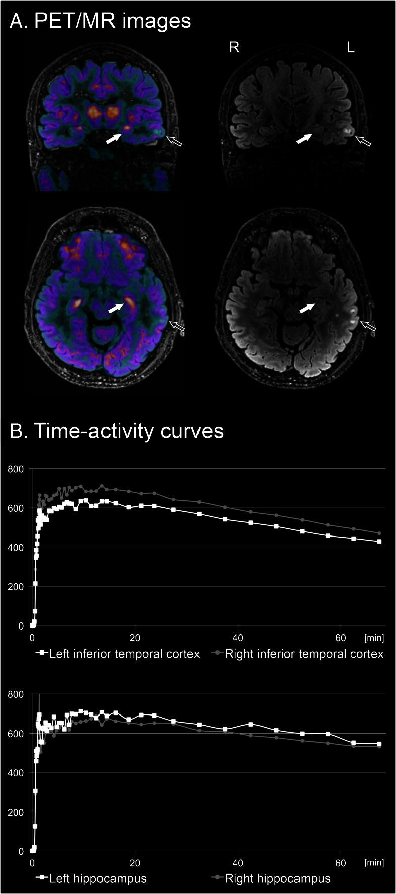
Fig. 1.
[18F]GE-179 PET/MR scan in a 57-year old temporal lobe epilepsy patient with signal abnormality in left inferior temporal cortex. a PET/MR fusion image (left) and MR FLAIR sequence (right) displaying parts of the lesion (empty arrow) and the hippocampus (full arrow). b Bilateral comparison of time-activity curves (TACs) in the inferior temporal cortex (above) and the hippocampus (below). Both visual analysis and TACs show reduced tracer uptake in the lesioned temporal cortex and slightly increased uptake in the ipsilateral hippocampus. Hypothetically, the extralesional increase of NMDA-receptor activation in the ipsilateral hippocampus could point to ongoing epileptogenesis and prospective studies will be needed to prove this assumption

