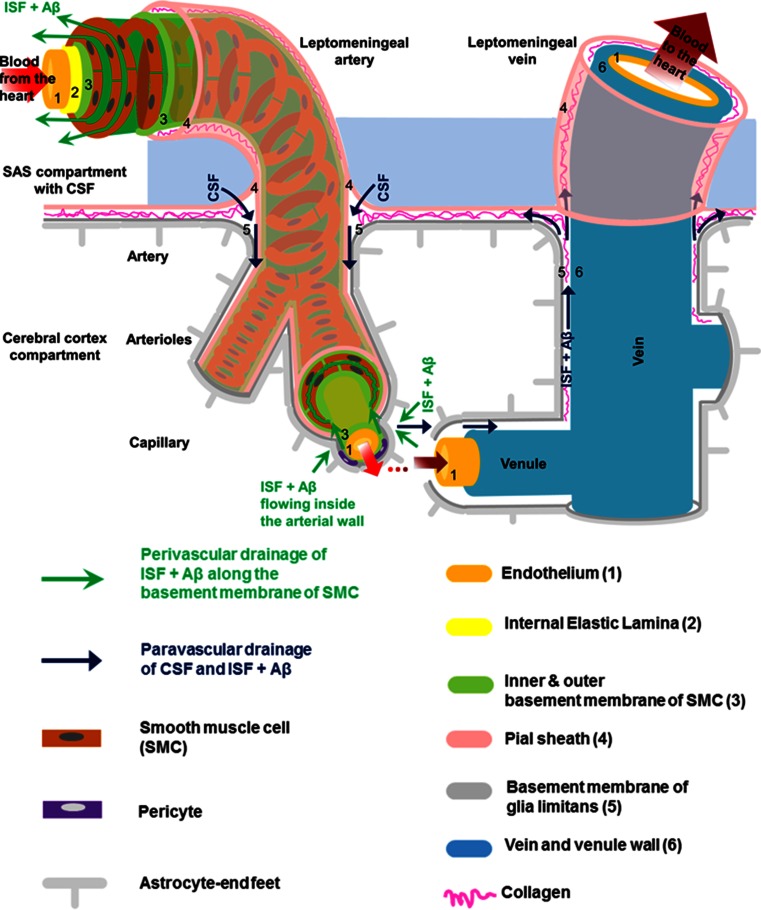
Fig. 3.
Schematic representation of the peri- and para-vascular spaces in the human cerebral cortex (not to scale). The subarachnoid space (SAS), filled with cerebrospinal fluid (CSF), is separated from the subpial space by the pia matter (Alcolado et al. 1988). A leptomeningeal sheath (the pial sheath shown in light pink) derived from the pia matter is also reflected on the walls of both arteries and veins that are crossing the SAS, thus separating the SAS from the cerebral cortex. Bundles of collagen are interposed in the subpial space and form the adventitia of leptomeningeal blood vessels, which is an expandable perivascular space. The pial sheath surrounding the leptomeningeal arteries in SAS continues to coat the arteries as they penetrate the cortex and it is closely applied to the outer basement membrane of smooth muscle cells (SMCs). Nonetheless, the veins from the cerebral cortex do not have a coating pial sheath. The middle layers of basement membrane (dark green), situated between layers of SMCs, represent the pathway for the perivascular drainage of solutes out of the brain. As emphasized, solutes do not drain along the inner or outer basement membrane layers of SMCs (light green). For the sake of simplicity, we illustrated only one layer of SMCs spiralling within the arterial wall. The perivascular drainage pathway (green arrows) follows a similar spiral trajectory which is spanned by a matrix of proteins. The basement membrane of the glia limitans (dark grey) is closely applied to the pia matter as the artery enters the brain. Thus another narrow perivascular space is created between the irregular surface of the pial sheath and the outer basement membrane of the SMCs and that of the glia limitans, respectively (Zhang et al. 1990). The narrow perivascular space has also been presented under the nomenclature of “paravascular space” and it has been proposed to be part of the glymphatic pathway (dark blue arrows). The cortical para-arterial pathway was referred to as the Virchow–Robin space (VRS). However, in the era of electron microscopy the exact boundaries of the VRS are not clearly defined (Color figure online)

