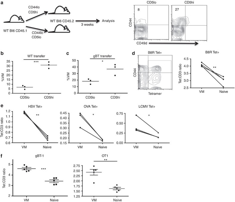
Figure 2. CD5 expression is a predictor of a naive cell's propensity to become VM.
(a) Naive (CD44lo), CD5hi (the 20% highest CD5 expressers) or CD5lo (the 20% lowest CD5 expressers) CD8 T cells were sorted out of unmanipulated WT mice, and 4.0 × 10⁵ of each population were adoptively transferred into congenic WT recipients, which were left undisturbed for 3 weeks (left). Recipients were then killed and splenocytes were analysed for transferred cells changing phenotype (right, per cent VM in quadrant). (b,c) Comparisons between WT mice receiving WT (b) or gBTxRag−/− (c) transferred cells (*P<0.05, ***P<0.001, t-test; n=3 per group). (d,e) Tetramer staining and pulldown of CD8s specific for foreign cognate antigen in WT mice. Representative contour flow plot of CD44/tetramer staining for B8R tetramer (left), overlay of pseudocolour dot plots for CD3 and tetramer staining in naive (blue) and VM (red) CD8 T cells. Comparison within mice of tetramer:CD3 gMFI staining on individual tetramer+ (Tet+) cells (d, B8R [TSYKFESV]; e, HSV [SSIEFARL], OVA [SIINFEKL], LCMV [KAVYNFATC]; *P<0.05, **P<0.01, paired t-test, representative of 2–4 experiments with n=3 per group). (f) PBMCs from gBT-1 and OTI TCR transgenic mice were stained with the HSV and OVA MHCI tetramers, respectively, as described above. VM and naive populations were compared within mice for Tet:CD3 ratios (**P<0.01, ***P<0.001, paired t-test; n=5 per group, error bars represent s.e.m.).

