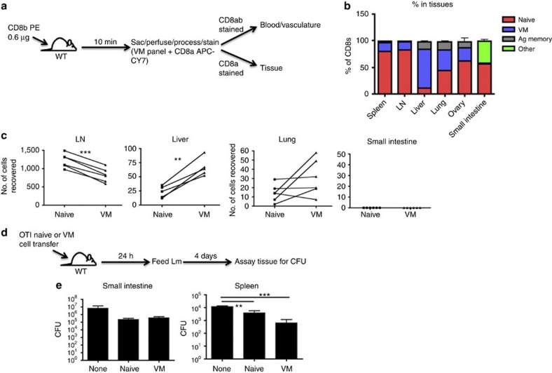
Figure 6. VM cells show a tropism for certain tissues under steady-state and trafficking conditions.
(a,b) WT mice were injected with CD8b antibody that was allowed to briefly circulate. Mice were then killed and stained with an antibody panel to determine naive, VM and antigen-experienced CD8 T cells, and whether the cells were in the vasculature or tissues (a). (b) Representative bar graphs of T-cell distribution (pooled data from two experiments with n=3 per experiment, error bars represent s.e.m.). (c) WT splenocytes were sorted into CD8 naive and VM populations, stained with different tracking dyes and co-adoptively transferred into recipient WT congenic mice. Recipients were killed 48 h later and tissues were processed as in a, and transferred cell populations were compared (**P<0.01, ***P<0.001, paired t-test, data combined from two experiments with n=3 per group). (d) OTI splenocytes were sorted into naive and VM populations and adoptively transferred into WT mice. One day later, mice were orally challenged with Lm expressing mutant internalin-A, and killed 4 days subsequent to challenge. (e) Tissues of mice orally infected were assayed for Lm CFUs (**P<0.01, ***P<0.001, one-way analysis of variance, representative of two experiments with n=3 per group, error bars represent s.e.m.).

