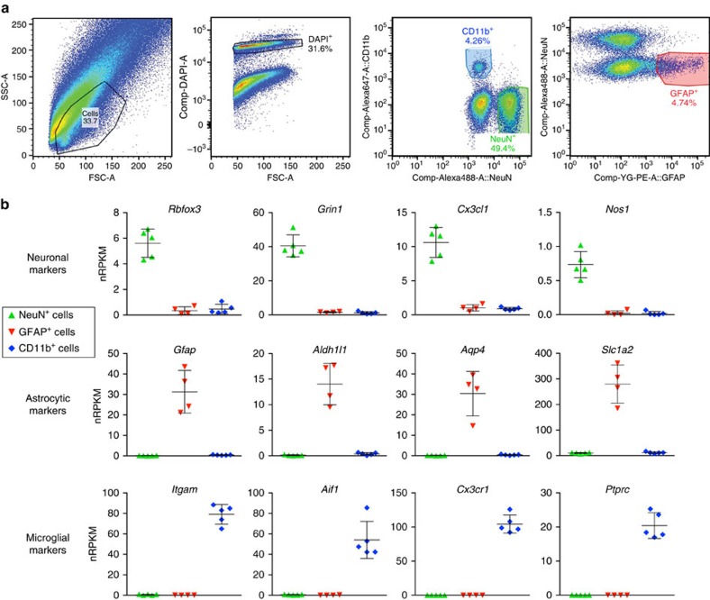
Figure 4. Antibody-based FACS and RNA-Seq for CNS cell types from adult brain tissue.
(a) Left panel shows forward/side scatter (FSC/SSC) of dissociated cortical tissue. DAPI+ events (‘cells') were gated to select for nuclei-containing singlets (second panel), and neurons and microglia were isolated as two distinct populations corresponding to NeuN+CD11b– and NeuN−CD11b+ cells, respectively (third panel). The NeuN− population was further used to isolate GFAP+ astrocytes (last panel). (b) RNA-Seq data confirmed that NeuN+ sorting enriched for cells expressing neuronal markers, GFAP+ sorting enriched for cells expressing astrocytic markers, and CD11b+ sorting enriched for cells expressing microglial markers (n=5 animals; one astrocyte sample was excluded from the analysis due to evidence of neuronal contamination). Bars represent mean±s.d. (Prism).

