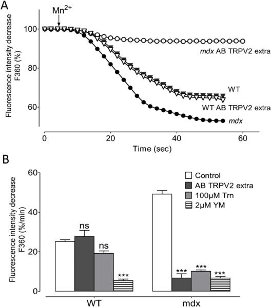
Fig. 3. Influx in stretched cardiomyocytes in presence of TRPV2 channels antibodies.
(A) example of recordings of fura-2 fluorescence during perfusion with 300 μM Mn2+ obtained from stretched WT (black triangles) and stretched mdx (black circles) and after incubation with antibody against TRPV2 (AB TRPV2 extra) from stretched WT (open triangles) and mdx (open circles) cardiomyocytes. (B) Effect of inhibitors on manganese quench rate in stretched cardiomyocytes. Stretched cells were pre-incubated with antibody against an extracellular epitope of TRPV2 (dark grey bars), 100 μM tranilast (clear grey bars) and 2 μM YM-48483 (horizontal hatching bars) in stretched WT and mdx cardiomyocytes. Open bars represent the control. Measurements are represented as slopes of the Mn2+-induced decreasing phase of fura-2 fluorescence and expressed as percent of decrease per minute. Bar graphs represent maximal rates of fluorescence decrease induced by Mn2+ ± SEM. *** P < 0.001; ns, not significant.

