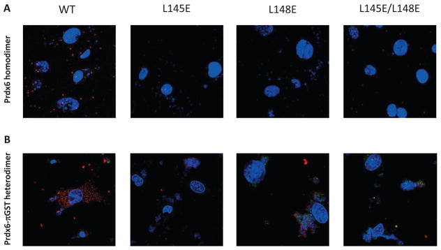
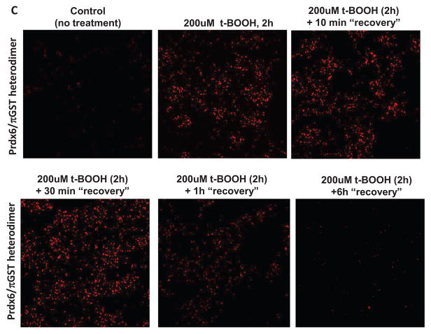
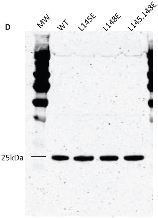
Figure 6. Effect of mutation on the dimerization of Prdx6 determined by the Proximity Ligation Assay (Duolink) test.
Prdx6 null mouse pulmonary microvascular endothelial cells expressing WT or mutant Prdx6 were fixed and probed by the DuoLink reaction according to the manufacturer’s protocol. Cells in (A) and (B) were counter-stained with 4′, 6–diamidino-2-phenylindole (DAPI) to show nuclei. Images in (A), (B), and (C) are representative of 3 or more fields that were examined for each of n=3 independent incubations. (A) Assay for homodimerization. Both arms of the DuoLink probe were conjugated to the primary antibody against Prdx6. Red fluorescence indicates proximity of two Prdx6 monomers. (B) Assay for heterodimerization. Cells were incubated with 200 μM t-BOOH for 2 h, and then processed using primary antibodies against Prdx6 and πGST for the 2 arms of the DuoLink probe. Red fluorescence indicates proximity of the Prdx6 and πGST monomers. (C) Reversibility of Prdx6: πGST heterodimerization. Experimental conditions as in (B). Panels show control (no t-BOOH), treatment with 200 μM t-BOOH for 2 h, and “recovery” for 10 min, 30 min, 60 min, or 6 h after removal of t-BOOH from the incubation medium. (D) Immunoblot analysis. Western blot of wild type (WT) and mutant (L145E, L148E, L145/148E) Prdx6 indicating that the mutant proteins react with the anti-Prdx6 antibody. MW, molecular mass markers.


