
Figure 2.
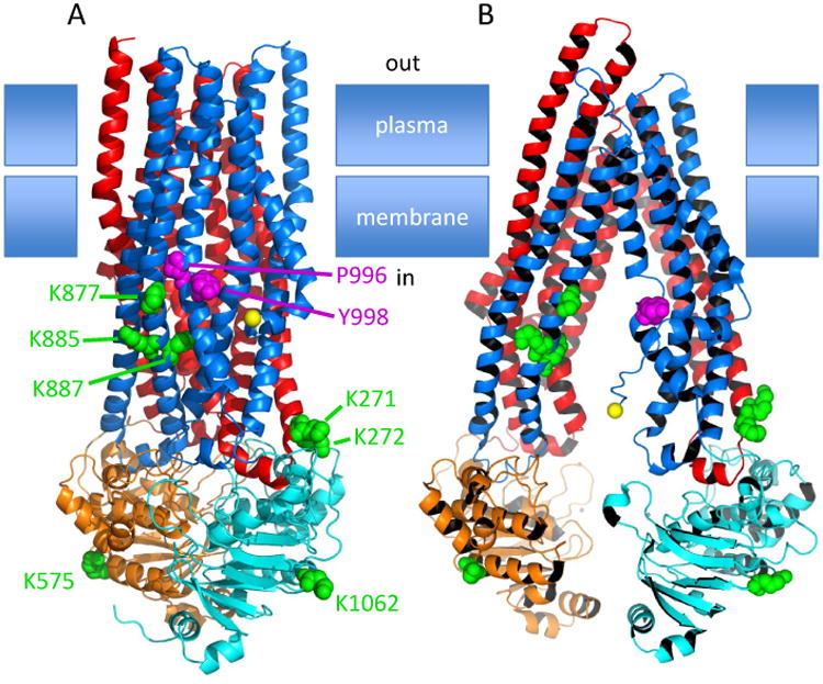
Cartoon representation of two conformations of ABCB1. (A) Homology model based on Sav1866 (pdb 2HYD) from S. aureus. (B) Model of mouse Abcb1a (pbd 35GU). In each case the domains are coloured from the amino terminus as follows TMD1, red; NBD1, orange; TMD2, blue; NBD2, cyan. The side chains of the putative NEDD4-1 binding motif (PxY) are shown as magenta spheres (P996 and Y998). The ubiquitinated lysines are shown as green spheres. The linker region linking NBD1 to TMD2 could not be modelled in either conformation therefore the precise position of K685 is not known but must be close to the first resolved residue in the amino-terminal region of TMD2, which in the closed conformation is W698, and in the open conformation is equivalent to L688 of human ABCB1. The C-alphas of W698 and the equivalent of L688 are shown as yellow spheres in the blue domain.