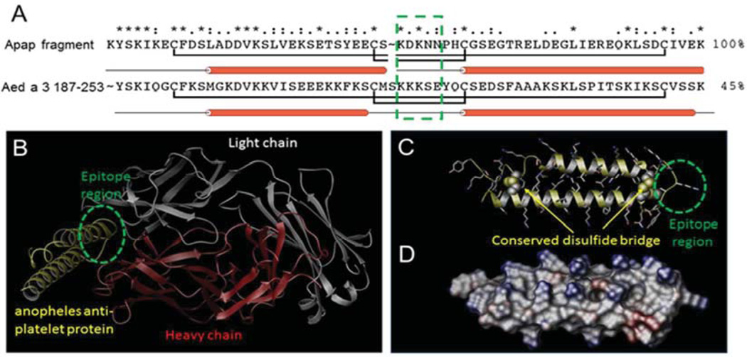
Figure 1.
Homology model of the Aed a 3 C-terminal region. (A) Sequence alignment between the apap fragment and Aed a 3 C-terminal region. The two conserved disulfide bridges are highlighted with the antigenic epitope region boxed in green. (B) X-ray structure of apap fragment (yellow) in complex with mouse FAB antibody consisting of a light chain (white) and a heavy chain (red). (C) Homology model of the Aed3 C-terminal region 187-253 with structurally conserved disulfide bridges. The antigenic epitope region is highlighted in green. (D) Electrostatic potential surface model of the Aed a 3 C-terminal region with the positively- and negatively-charged surfaces in blue and red, respectively.

