Fig. 2.
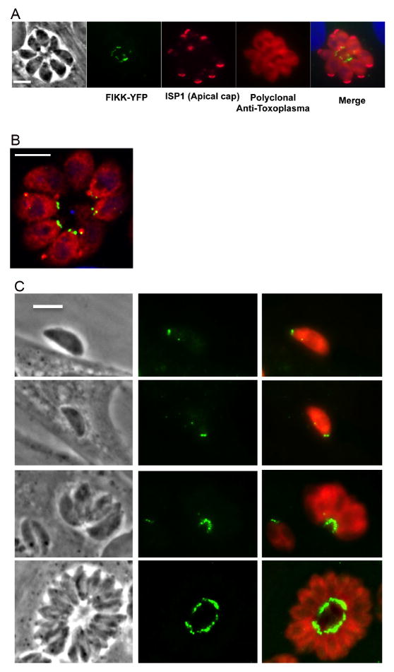
Immune-localization of Toxoplasma gondii TgFIKK kinase endogenously tagged with Yellow Fluorescent Protein (YFP). YFP-tagged protein (YFP-FIKK) was exclusively localized to the posterior end of the parasite throughout the parasite’s lytic cycle. (A) IIFA showing the YFP-tagged FIKK protein (green) in the posterior end of the parasite infecting human foreskin fibroblast (HFF) cells at 24 h p.i. The apical cap portion of the parasite was stained with a monoclonal antibody (mAb) to apical protein inner membrane complex (IMC) subcompartment protein-1 (ISP1) and is shown in pink. Polyclonal rabbit anti Toxoplasma antibody was used to stain for parasites (red). The host nucleus is stained with DAPI (blue). Scale bar = 5 μm. (B) Zeiss LSM 800 with Airyscan image of the YFP-tagged FIKK protein 24 h p.i. The FIKK protein was localized to two adjacent punctate dots in the basal end of the parasite (green). Parasites (red) are stained with a polyclonal antiserum to the parasite. Scale bar = 5 μm. (C) Localization of the FIKK protein during the parasite’s lytic cycle. The FIKK protein was constitutively and exclusively expressed in the basal end of tachyzoites throughout the parasite’s lytic cycle. IFA of the YFP tagged FIKK protein (green) in a parasite during invasion and at 2 and 36 h p.i. Localization of the FIKK kinase at 24 h p.i. is shown in Figs. 4A, C. Scale bar = 5 μm.

