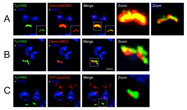
Fig. 3.
Co-localization analysis of the Toxoplasma gondii N-terminal Ty-tagged FIKK protein with markers of the basal complex. FIKK protein localizes in the basal complex. (A) N-terminal Ty-tagged FIKK co-localizes with membrane occupation and recognition nexus protein (MORN1) inside the basal complex. (B) The FIKK protein sits apical to IMC5. (C) The FIKK protein sits apical to the posterior cup marker Centrin2. Ty-tagged FIKK is pseudo-colored green. MORN1, IMC5 and Centrin2 are pseudo-colored red. Magnification of boxed areas is shown in the zoom. Scale bar = 2 μm.

