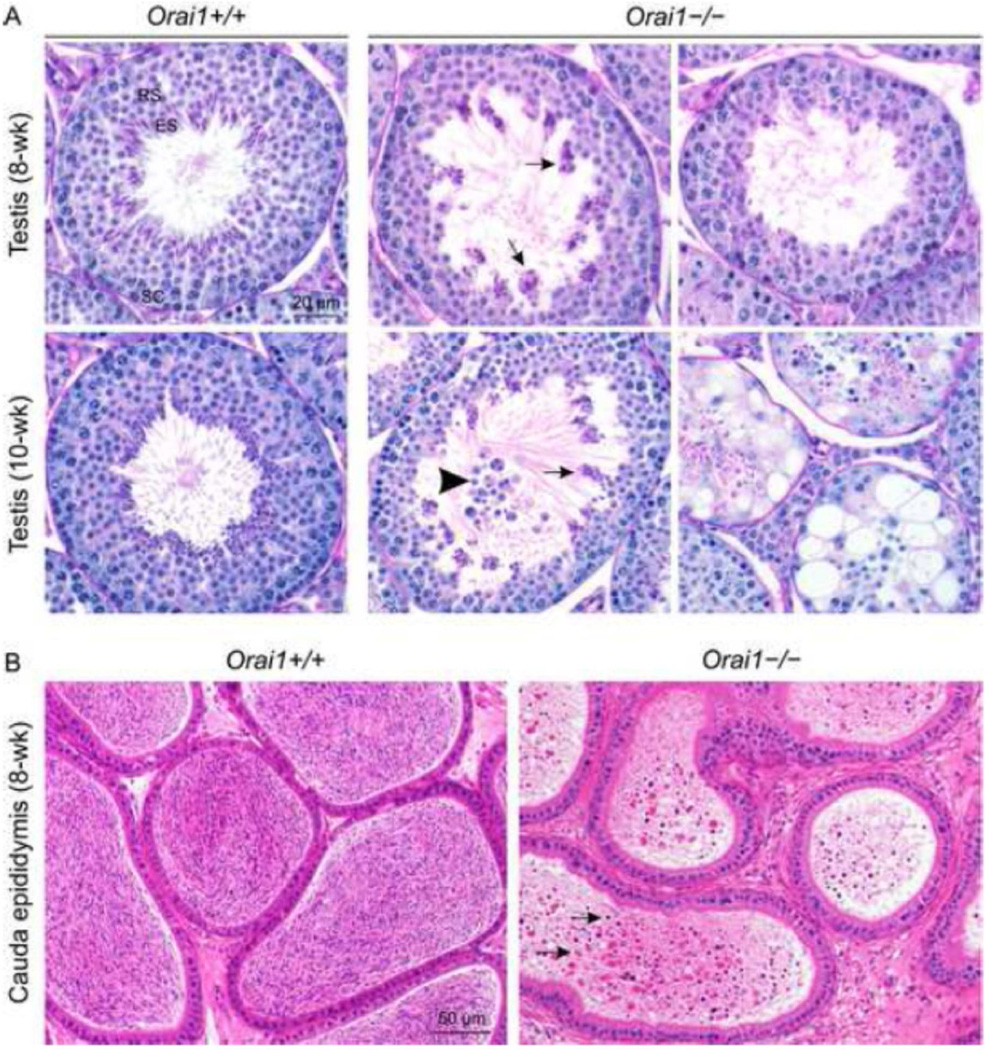
Fig. 6.
Histopathological analysis of the testis and cauda epididymis of Orai1−/− mice. A, Stage VII seminiferous tubules from 8- and 10-week-old Orai1+/+ mice with elongated spermatids lining the lumen of the seminiferous tubule (Periodic Acid Schiff-Hematoxylin staining; RS, round spermatids; ES elongated spermatids; SC, spermatocytes) versus Orai1−/− mice with numerous poorly populated seminiferous tubules. Arrows: degenerate and clumped elongated spermatids. Arrowhead: exfoliation of round spermatids. B, Hematoxylin and eosin staining of the cauda epididymis from 8-week-old Orai1−/− versus Orai1+/+ mice. The cauda epididymis in WT mice is full of normal sperm, whilst that of Orai1−/− mice contains very low number of sperm admixed with cellular debris (arrows). For 8-week-old samples (n = 3 mice/group) and for 10-week-old samples (n = 2 mice/group).

