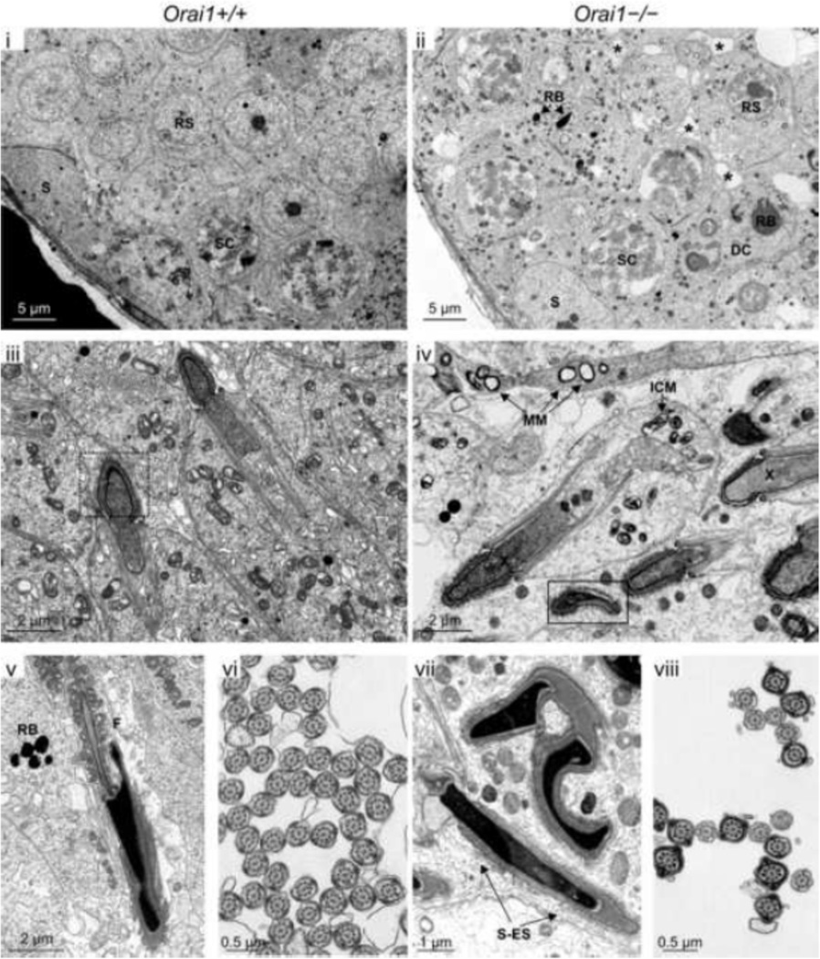
Fig 8.
Ultrastructural analysis of randomly selected seminiferous tubules within the testis of Orai1+/+ and Orai1−/− mice. Digital transmission electron photomicrographs of seminiferous tubules from Orai1+/+ and Orai1−/− mice were compared. Individual letters on each image identify the particular structure: DC, degenerating cell; F, flagella; ICM, intracristal mitochondrial swelling; MM, matrical mitochondrial swelling; RB, residual body; RS, round spermatid, S, Sertoli cell; SC, spermatocyte; and S-ES, Sertoli-elongating spermatid junction. Small asterisks in (ii) show swelling of the junctional complexes between spermatids and/or Sertoli and spermatids, whereas swelling along the junctional complexes were sparse in the Orai1+/+ tubules (i). Dashed box in (iii) shows a normal spermatid head at early elongating stage versus in the solid box (iv), early elongating spermatids had an abnormal head and loosely stippled to granular nuclear material (X). n = 3 mice/group. Lastly, when observed, distinct ultrastructural defects in later elongating stages included extensive malformations of the sperm head. (Fig. 7–iv, 7vii, 7-viii). Moreover, cross-sections of the flagella from a Orai1+/+ and Orai1−/− mouse when compared showed mostly normal formation; however, there was clearly a difference between the numbers of flagella in cross section from an Orai1+/+mice compared with Orai1−/− mice, the latter having a marked decrease in the number of cross-sectional flagella (Fig. 7–vi versus 7-viii).

