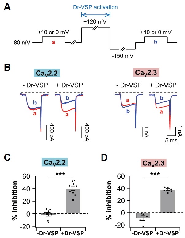
Fig. 3.

PI(4,5)P2 depletion by Dr-VSP decreases both CaV2.2 and CaV2.3 currents. TsA201 cells were co-transfected with Dr-VSP and either CaV2.2 or CaV2.3 channels. (A) Standard protocol for Dr-VSP activation. Cells received a test pulse (a), then a depolarization to +120 mV for 1 s to activate the Dr-VSP, a hyperpolarization to −150 mV for 0.4 s to remove the voltage-dependent inactivation, and a second test pulse (b). CaV2.2 or CaV2.3 currents were measured before and after the Dr-VSP activation at +10 mV or 0 mV, respectively. (B) Left, CaV2.2 current regulation by a membrane depolarization to +120 mV for 1 s in control (-Dr-VSP, n = 8) and cells expressing Dr-VSP (n = 9). Right, CaV2.3 current regulation in control (n = 6) and cells expressing Dr-VSP (n = 6). (C, D) Summary of % inhibition by Dr-VSP-induced PI(4,5)P2 depletion in CaV2.2 (C) and CaV2.3 (D) channels. Data are mean ± SEM. *** P < 0.001, compared with - Dr-VSP.
