
Fig. 4.
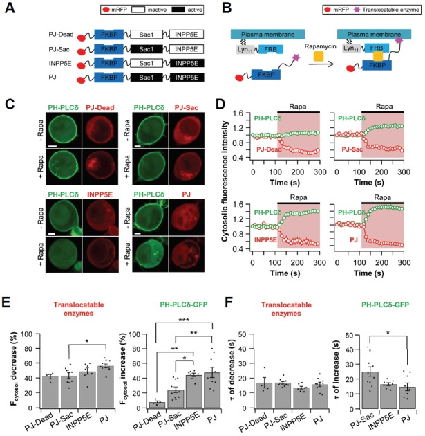
Plasma membrane PI(4,5)P2 levels are reduced by translocation of PJ-Sac, INPP5E, and PJ. TsA201 cells were co-transfected with Lyn11-FRB, PH-PLCδ-GFP, and one of the following four constructs: PJ-Dead, PJ-Sac, INPP5E, or PJ. (A) Construct diagrams of the four translocatable phosphoinositide phosphatases. (B) Diagram of the chemically induced dimerization (CID) system. Rapamycin triggers the translocation of cytoplasmic phosphatases to the plasma membrane. (C) Confocal images of cells expressing PJ-Dead (upper left), PJ-Sac (upper right), INPP5E (lower left), or PJ (lower right) with PH-PLCδ-GFP. Images are from before (Upper) and after (Lower) the application of rapamycin (1 μM) for 180 s (Scale bar, 5 μm). (D) Relative cytosolic intensity of PH-PLCδ-GFP (Green) and translocatable enzyme (Red) for the cells in (C). (E) Summary graph of % decrease in cytosolic translocatable enzymes and of % increase in cytosolic PH-PLCδ-GFP by addition of rapamycin (n = 4 for PJ-Dead; n = 10 for PJ-Sac; n = 7 for INPP5E; and n = 9 for PJ). (F) Summary graph of the time constant for rapamycin-induced decrease in cytosolic translocatable enzymes and for rapamycin-induced increase in cytosolic PH-PLCδ-GFP (n = 4 for PJ-Dead; n = 10 for PJ-Sac; n = 7 for INPP5E; and n = 9 for PJ). * P < 0.05, ** P < 0.01, and *** P < 0.001, with one-way ANOVA followed by Bonferroni post-hoc test.