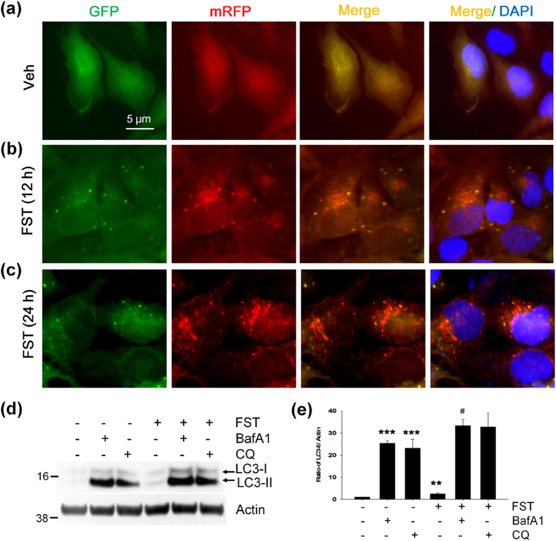
Figure 8. Fisetin increases autophagy flux.
(a–c) Human neuronal H4 stable cells stably expressing mRFP-GFP-LC3 were treated with either DMSO (Veh) or 10 μM fisetin (FST). The cells were fixed with 4% paraformaldehyde, and fluorescence signals were observed under the epifluorescence microscope. (d) Mouse cortical cells (T4) were treated with either DMSO (Veh) or 5 μM fisetin (FST) for 12 h. The cells were then incubated for an additional 18 h following treatment with 100 nM bafilomycin A1 (Baf A1) or 50 μM chloroquine (CQ). The level of LC3-II was analyzed by immunoblotting using the anti-LC3 antibody. (e) Bar graphs represent the relative ratio of LC3-II normalized with that of actin. Data shown are mean ± SE of three independent experiments and were analyzed using Student’s t test. (**p < 0.01; ***p < 0.001; *cells treated with Baf A1, CQ or fisetin versus cells not treated), (#p < 0.05; #cells treated with fisetin plus Baf A1 versus cells treated with Baf A1 only).

