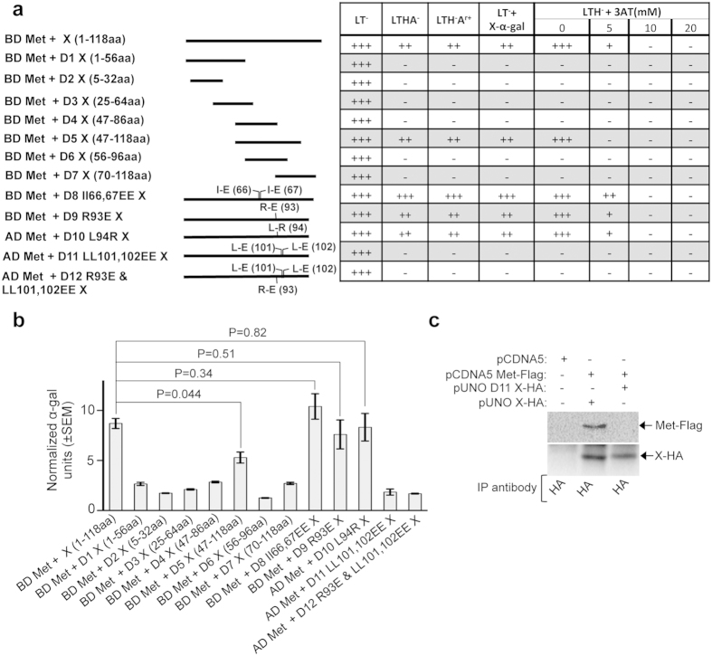
Figure 4. Deletion and point mutation analyses of the X-domain residues responsible for its interaction with the methyltransferase protein.
(a) Y2H analyses of the interaction potential of various deletion and point mutants of X-domain with full-length methyltransferase (Met). Length of the deletion mutants is graphically represented as solid lines and amino acid position of point mutants are indicated in brackets. AD: Activation domain, BD: Binding domain, +++: Strong growth, ++: Moderate growth, +: Poor growth, −: No growth, L: Leucine, T: Tryptophan, H: Histidine, A: Adenine hemisulfate, Ar: Aureobasidin, “−”: Deficiency in the medium, “+”: Supplemented in the medium. (b) Quantitative α-galactosidase assay of Y2H gold transformants represented in Fig. 4a. Normalized α-galactosidase units are plotted as ± SEM of triplicate samples. (c) CoIP of Huh7 cell extract expressing empty vector or wild type X (X-HA) and methyltransferase (Met-Flag) or LL-EE mutant X (D11 X-HA) and methyltransferase (Met-Flag), immunoprecipitated using anti-HA antibody and immunoblotted using anti-Flag (upper panel) or anti-HA (lower panel) antibodies.

