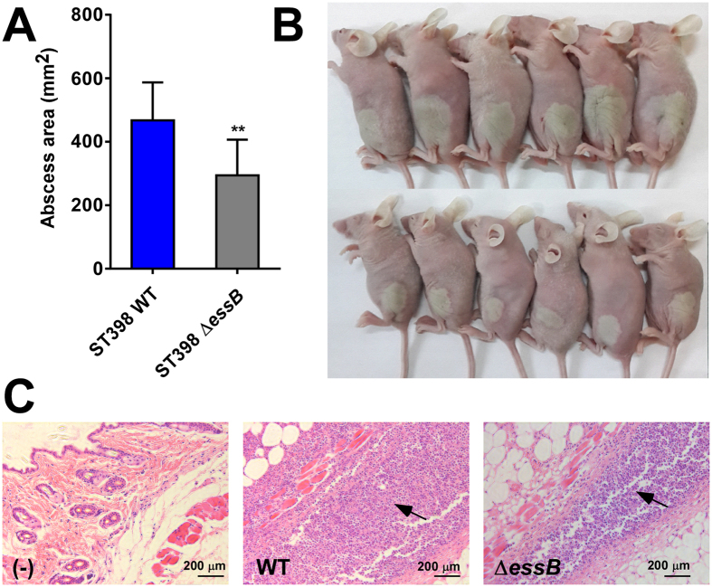
Figure 3. Role of ESS in CA-SA ST398 skin infection.
Mouse abscess model with essB deletion mutant versus isogenic wild-type representative ST398 isolate. Number of mice used: n = 11/group. (A) Abscess areas on day 2 after infection. **p < 0.01 (unpaired t-test). (B) Representative abscesses on day 2 after infection. (C) H&E staining of abscess skin tissue harvested on day 4. (–), control without challenge. Note considerable infiltration of inflammatory cells and damage of the subcutaneous structure in the wild-type and essB mutant samples, noticeably stronger in the wild-type sample (arrows).

