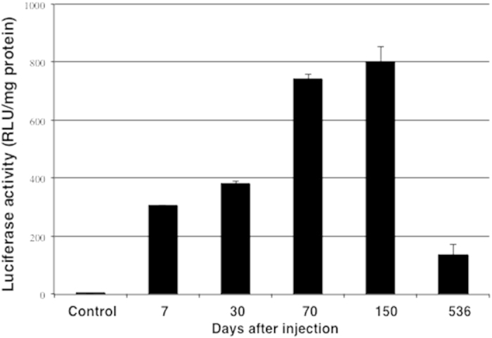
Figure 2. Luminescence quantification of luciferase activity in Atlantic salmon muscle tissue 7, 30, 70, 150 and 536 days after intramuscular injection of pGL3-35S.

PBS injected fish served as control. Each bar represents the mean luciferase activity of five fish and T-bars represent the S.D. The measured luciferase activity was normalised for total protein (mg).
