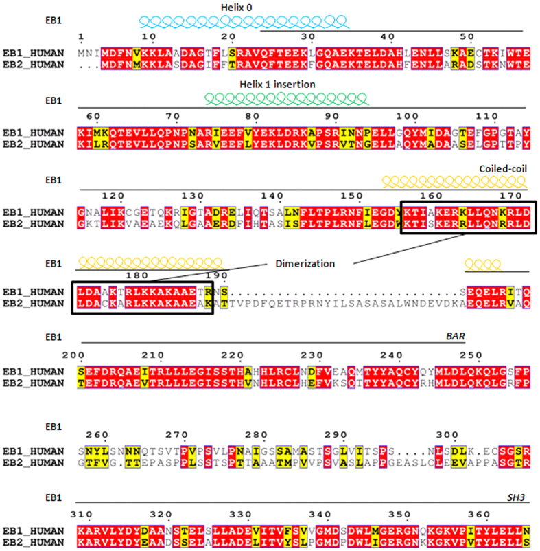
Figure 1. Schematic alignment of the amino acid sequences of human EB1 and EB2.
Alignment of protein sequences is exhibited using ESPript 3.0. Conserved residues (red) and similar residues (yellow) are indicated. The amphipathic helix 0 (light blue spirals) that precedes the BAR domain, and the helix 1 insertion (green spirals) were conserved between EB1 and EB2, whereas the coiled-coil domains (yellow spirals) were not conserved. Dimer motifs in the coiled-coil domain are marked with black boxes.

