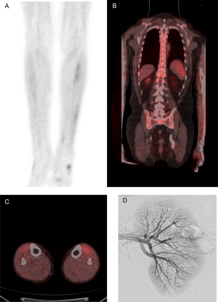
Figure 3:
Radiological findings illustrating bone marrow uptake and organ involvement. [18F] Fluorodeoxyglucose positron emission tomography (FDG-PET) scan demonstrating increased uptake of FDG in the (A&C) calf musculature and (B) bone marrow of the axial skeleton, extending into the proximal humeri. (D) Digital subtraction imaging showing microaneurysms.

