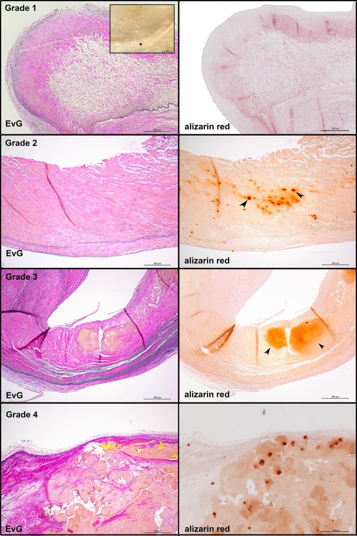
Figure 1.

General morphological findings by grading aortic valve calcification and structural damage according to Warren and Yong.17 Left panels, van Gieson's staining demonstrating the 3 anatomic layers: fibrosa, spongiosa, and ventricularis. The fibrosa with the aortic side of the valve is to the top. Right panels, adjacent sections stained with alizarin red S to depict calcified areas. First panel, grade 1 (inset: macroscopic appearance of a representative early valvular cusp lesion. The valvular cusp was carefully inspected under a dissection microscope, and areas characterized by focal yellowish patches or streaks [asterisks] were defined as grade 1 lesions). Second panel, grade 2. Third panel, grade 3. Fourth panel, grade 4. EvG indicates Elastica van Gieson.
