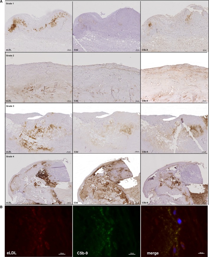
Figure 3.

Colocalization of eLDL with C3d and C5b‐9. A, Sequential sections of the 4 grades of aortic valve calcification and structural damage stained for eLDL (left panels), C3d (middle panels), and C5b‐9 (right panels). Note the close intermingling and overlap of the different antigens in all of the 4 grades. In all panels, the fibrosa with the aortic side of the valve is to the top. B, Representative double immunofluorescence staining of another early valvular cusp lesion demonstrating perfect overlap of eLDL and C5b‐9 (merge, yellow indicates colocalization). The aortic side of the valve is to the right. eLDL indicates enzymatically modified low‐density lipoprotein.
