
Figure 1.
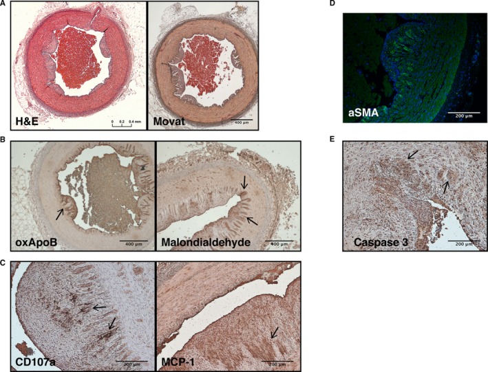
Representative histology of atherosclerotic lesions in the coronary arteries of adult RFH swine. A, Hematoxylin and eosin (H&E) staining and Movat's Pentachrome revealed wall thickening, fibrin accumulation, and plaque buildup in coronary arteries. B, α‐SMA‐positive cells were detected in the subendothelial region. C, Lipid oxidation was observed by immunostaining of oxApoB and malondialdehyde. D, Apoptotic cells were identified throughout the arteries by immunostaining for caspase 3. E, Leukocyte degranulation (CD107a) and the secretion of MCP‐1 were also observed in the intima, as expected in an atherosclerotic plaque. Scale bars: 400 μm (A and C); 200 μm (B, D, and E). Arrows indicate areas representative of positive staining. N=4 animals. MCP‐1 indicates monocyte chemoattractant protein‐1; oxApoB, oxidatively modified apolipoprotein B; RFH, Rapacz familial hypercholesterolemic; α‐SMA, alpha‐smooth muscle actin.