
Fig. 5.
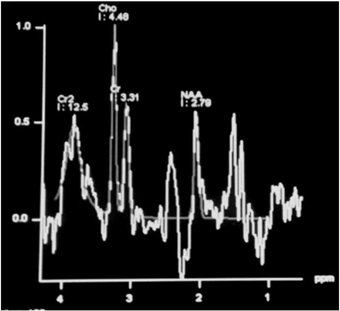
H1MRS spectra acquired at low TE (30ms) shows an elevated alanine peak at 1.48 ppm proposing a possibility of meningioma. This is the first peak seen in the spectra (after lipid at 1.3) following which the NAA peak is reduced as meningioma is an extra-axial tumor (the NAA seen is due to contamination from adjacent brain). Note the prominent choline peak with a small creatine peak, which signifies a hyppometabolic tumor.