
Figure 1.
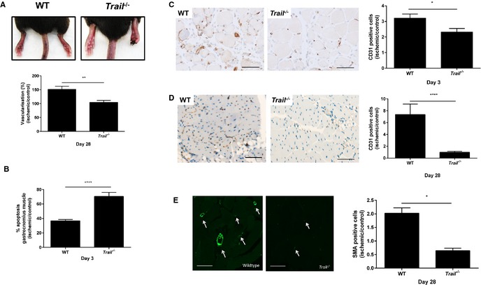
Trail−/− mice have reduced vascularity following hindlimb ischemia. A, WT and Trail −/− mice hindlimb after hindlimb ischemia. Top panel, representative images showing necrotic toes of Trail −/− mice at 14 days. Bottom panel, vascularization of the ischemic and nonischemic hindlimbs were evaluated by real‐time in vivo 3‐dimensional Vevo ultrasound imaging using contrast agents (Vevo MicroMarker) at 28 days (WT, n=5; Trail −/−, n=5). B, Apoptosis was assessed using the Cell Death Detection ELISA from muscle tissue 3 days after hindlimb ischemia was induced; % apoptosis in ischemic muscle over control muscle (WT, n=3; Trail −/−, n=3). CD31 staining in gastrocnemius muscle at (C) 3 days (WT, n=5; Trail −/−, n=4) and (D) 28 days postischemia (WT, n=5; Trail −/−, n=5). E, SMA staining at 28 days postischemia. Left panel shows staining from ischemic leg. Right panel shows quantification (WT, n=5; Trail −/−, n=4). Data were normalized to the nonischemic control leg. Magnification bars=50 μm. Results are mean±SEM; Mann–Whitney U‐test; *P<0.05, **P<0.01, and ****P<0.0001. SMA indicates smooth muscle α‐actin; WT, wildtype.