Figure 7.

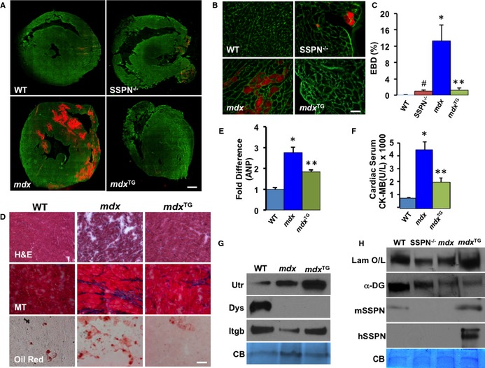
SSPN attenuates membrane damage in DMD hearts. A, Mice were injected with EBD to detect regions of membrane instability (visualized by red fluorescence). Transverse cryosections of heart muscle from 12‐month‐old WT, SSPN‐null (SSPN −/−), DMD model (mdx), and mdx:SSPN‐Tg (mdx TG) mice were stained with laminin antibodies (green) to visualize individual myofibers. Bar, 900 μm. B, Magnified (×40) images of representative sections from (A). Bar, 50 μm. C, Quantification of EBD‐positive fibers (n=3 hearts per genotype). D, Cardiac muscle from 4‐month‐old WT, mdx and mdx TG mice were stained with H&E, MT, and Oil Red (n=2). Bar, 50 μm. E, Relative changes in gene expression changes of ANP were assessed by qRT‐PCR in 4‐month‐old WT, mdx, and mdx TG hearts and number of mice utilized was (WT (n=4), mdx (n=5), mdx TG (n=5). F, Serum CK‐MB levels were detected in WT; mdx and mdx TG groups of mice (WT (n=5), mdx (n=6), mdx TG (n=5). G, Immunoblots of sWGA cardiac eluates show changes in key proteins in WT, mdx and mdx TG hearts (n=3 experiments, samples from 3 different mice of each genotype) whereas, in (H) Immunoblots show changes in laminin binding in a laminin overlay experiment (Laminin O/L) upon overexpression of SSPN in WT, mdx and mdx TG hearts (n=3 experiments, samples from 3 different mice of each genotype). Equal sWGA protein samples (30 μg) were resolved by SDS‐PAGE and immunoblotted with the indicated antibodies. CB staining is shown as a loading control. All data are presented as averages and error bars are expressed as SEM. Statistics were performed by 1‐way ANOVA with Bonferroni correction for individual groups in (C through F) and *P‐values ≤0.05 are indicated. For (C) *P=0.018 mdx vs WT, **P=0.027 mdx vs mdx TG, # P<0.001 SSPN −/− vs WT; (E) *P=0.003 WT vs mdx, **P=0.036 mdx vs mdx TG and (F) * P<0.001 mdx vs WT and **P=0.009 mdx vs mdx TG. ANP indicates atrial natriuretic peptide; CB, Coomassie blue; CK‐MB, cardiac creatine kinase; DMD, Duchenne muscular dystrophy; EBD, Evans blue dye; H&E, hematoxylin & eosin; mdx, Duchenne muscular dystrophy mouse model; mdx TG, mdx mice overexpressing human sarcospan; MT, Masson's trichrome; qRT‐PCR indicates quantitative reverse transcription–polymerase chain reaction; SDS‐PAGE, sodium dodecyl sulfate–polyacrylamide gel electrophoresis; SSPN, sarcospan; sWGA, succinylated wheat germ agglutin; WT, wild‐type.
