Figure 4.

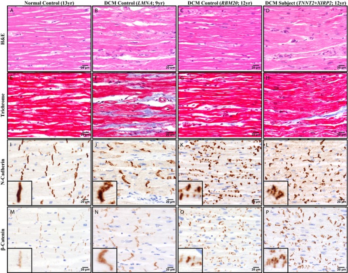
Histochemistry and immunohistochemistry revealed abnormalities in ICD structure in pediatric DCM. Representative photomicrographs of left ventricular myocardial tissue from a normal male control, age 13 (A, E, I, and M); a DCM female control harboring a LMNA mutation, age 9 (B, F, J, and N); a DCM female control harboring a RBM20 mutation, age 12 (C, G, K, and O); and the female DCM patient harboring TNNT2 and XIRP2 mutations, age 12 (D, H, L, and P). All 3 DCM patients showed nonspecific alterations (B through D, H&E=hematoxylin & eosin; F through H, Masson trichrome). N‐cadherin (I through L) and β‐catenin (M through P) immunohistochemistry revealed ICD abnormalities that differed among the DCM patients. The LMNA DCM control showed many irregular ICDs oriented obliquely, but with ICD length relatively preserved (J and N). In contrast, the RBM20 DCM control showed many short ICDs, including unusual linear arrays of multiple short parallel ICDs distributed along the sides of myocytes (K and O). The patient with TNNT2 and XIRP2 mutations showed severely disorganized ICDs, with numerous clustered groups composed of a mixture of oblique, short, and irregular forms (L and P). Immunoreactivity of ICDs for β‐catenin was increased in all DCM individuals relative to the normal control (M through P). Scale bars (A through P)=20 μm. DCM indicates dilated cardiomyopathy; ICD, intercalated disc.
