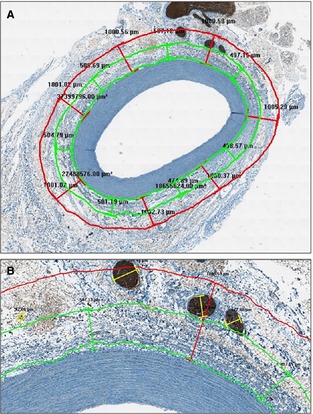
Figure 1.

Morphometric analysis of renal nerves. Measurements were related both to the entire area of the peri‐adventitial tissue and to 2 concentric rings that were located within 0.5 mm (called the “internal area”) and between 0.5 and 1 mm (defined as the “intermediate area”) from the beginning of adventitia (A, ×2). B, A higher magnification of (A) (×10).
