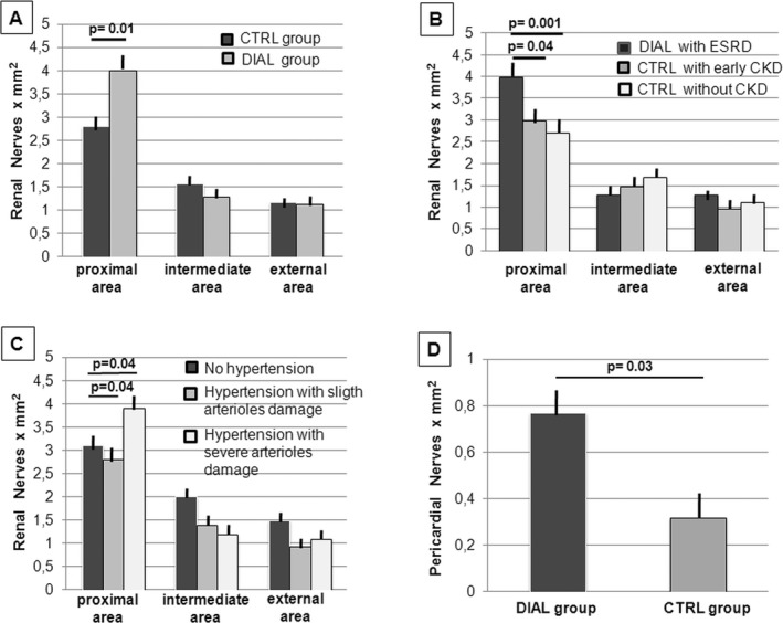
Figure 3.

Distribution of peri‐adventitial and pericardial nerves. A, Patients in the DIAL group showed a significant increase in nerve density in the internal area of the peri‐adventitial tissue compared with the CTRL group (P=0.01). No significant differences were observed in the intermediate and external areas. B, Patients in the CTRL group with chronic kidney diseases in an earlier stage of the disease showed a smaller number of nerve endings in the internal area of the adventitia compared with dialysis patients with ESRD (P=0.03) and no significant differences compared with CTRL patients with normal renal function (P=0.93). No significant differences were observed in the intermediate and external area. C, Regardless of dialysis, hypertensive patients with signs of severe arteriolar damage showed a nerve density in the internal area of the adventitia that was significantly higher than that of hypertensive patients with minimal arteriolar damage (P=0.04). In this last group, the nerve density was similar to that of normotensive patients. D, Patients of the DIAL group showed a significant increase in nerve density in the pericardium compared with the CTRL group (P=0.03). CKD indicates chronic kidney disease; CTRL, control; DIAL, dialysis; ESRD, end stage renal disease.
